VVector Bio offers excellent services for Adeno-associated virus (AAV), Lentivirus and Adenovirus production to support cell and gene therapy development
Sessions
1. Introduction to Adeno-Associated Virus
Adeno-associated virus (AAV) is a small, non-enveloped virus that is a member of the family of Parvoviridae – genus Dependoparvovirus – which means, it may depend on a helper virus to replicate. AAVs will only replicate if the cellular environment is altered dramatically, which may occur in the presence of a helper virus, genotoxic or DNA damaging compounds or metabolic inhibitors [15, 22]. However, replication without the presence of a helper virus has lower efficiency [6]. After infecting a host cell line, wild-type AAV genome persists as episomes or may be integrated into the host cell genome at low rates. Latent AAV will be rescued and activated upon subsequent helper virus infection, followed by replication and packaging of the viral genome[17].
AAV is not known to cause disease in humans, however precautions must be taken to avoid any kind of insertional mutagenesis. It is a well spread virus in nature, being 70-80% of the human population seropositive for anti-AAV antibodies against serotypes 1, 2, 3 or 5 [14].
Due to its safety profile and efficacy to deliver genes to dividing and non-dividing cells, AAV is one of the main viral vectors used in gene therapy, among others as retroviruses (RV), adenoviruses (AV) and lentiviruses (LV). Moreover, AAV is amenable to genetic engineering allowing rational design or molecular evolution of novel capsids with enhanced tissue specificity [4].
2. AAV in Gene Therapy
Recombinant AAV vectors have been used in clinical trials for diseases such as hemophilia, lipoprotein lipase deficiency, cystic fibrosis, spinal muscular atrophy, Leber’s congenital amaurosis, Parkinson’s disease, Pompe’s disease, Duchenne’s muscular dystrophy, among others [9].
In recent years, 3 AAV-based therapeutic products were approved: voretigene neparvovec (Luxturna®, Sparks Therapeutics), an AAV2 carrying the RPE65 gene for inherited retinal disease; onasemnogene abeparvovec-xioi (Zolgensma®, AveXis), an AAV9 carrying the human survival motor neuron (SMN) protein for spinal muscular atrophy; and alipogene tiparvovec (Glybera, UniQure), an AAV1 containing lipoprotein lipase gene for lipoprotein lipase deficiency [14, 32]. However, Glybera were discontinued for commercial reasons in 2017 [31]. Moreover, there are currently over 200 clinical trials ongoing [14].
3. AAV Biology
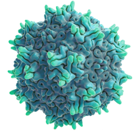
Adeno-associated virus (AAV) is a small, non-enveloped and single-stranded DNA (ssDNA) virus with a genome of 4.7 kb and 20-26 nm in diameter. The AAV genome contains two open reading frames (ORF) flanked by two 145 base inverted terminal repeats (ITRs) encoding replication proteins (Rep), capsid proteins (Cap), the assembly-activating protein (AAP) and the membrane-associated accessory protein (MAAP) [13].
The Inverted Terminal Repeats (ITRs) provide an origin of DNA replication by self-assembling into a T-shaped double-hairpin structure and are the only cis-acting elements required for genome replication and packaging. In wild-type AAV (wtAAV), this region flanks the two viral genes, rep and cap, responsible for virus replication, packaging and assembly. Also ITR sequences mediate AAV integration into target sequences within the host genome in the presence of Rep78, preferentially on a specific AAVS1 locus on chromosome 19 [14].
The rep gene encodes 4 non-structural Rep proteins that are responsible for AAV DNA replication, site-specific integration, transcription regulation and packaging of DNA into the capsids. Their apparent molecular masses consist of 78 kDa, 68 kDa, 52 kDa and 40 kDa, which designate the large (Rep78, Rep68) and small (Rep52, Rep40) Rep Proteins. The large rep proteins (Rep78 and Rep68) possess helicase, endonuclease and ATPase activities, essential for AAV DNA replication, and their expression is driven by p5 promoter. The small rep proteins (Rep52 and Rep40) are fundamental for virus packaging and their expression is driven by p19 promoter [19].
The AAV capsid is a 60-mer icosahedral virion consisting of three structural Cap proteins (VP1, VP2, and VP3) of 87 kDa, 73 kDa, and 62 kDa, respectively, that are expressed at an approximate 1:1:10 ratio. The cap gene expression is driven by the P40 promoter and is encoded by a single open reading frame regulated by alternative splicing [13].
In the same ORF of the cap gene is also encoded the non-structural Assembly-Activating Protein (AAP), a protein that is related to capsid assembly for most serotypes with the exclusion of AAV4, 5 and 11. Another protein, the Membrane-Associated Accessory Protein (MAAP) has recently been identified as a gene product in the VP1 region but its exact function needs to be elucidated [3].
4. AAV replication
AAV enters the cell upon attachment to a primary receptor, which varies through different serotypes, followed by endocytosis. AAV2, AAV3, and AAV6 attach to heparan sulfate proteoglycan (HSPG), while AAV1, AAV3, AAV4, and AAV6 attach to sialic acids and AAV9 to N-linked galactose. Another transmembrane protein named AAV receptor (AAVR) was identified as an essential protein for transduction of multiple AAV serotypes. After endocytosis, AAV is transported from early endosomes to the Golgi apparatus. Low pH in endosomal compartments and proteases trigger a conformational change in the AAV capsid, exposing the N-terminal domain of the VP1. This cap protein contains a phospholipase A2 domain (PLA2) and a nuclear localization signal, allowing the viral escape into the cytoplasm and the nuclear import. Once inside the nucleus, AAV is found in an episomal circular state during latency and integration may occur at very low rates (0.1-0.5%) [14].
Upon coinfection with a helper virus, transcription and replication of the AAV genome is initiated, starting the AAV lytic stage. For DNA replication, ITR acts as a primer to initiate second-strand synthesis resulting in a duplex structure. Then, Rep78 and Rep68 nick the terminal resolution site (trs) within the ITR sequence converting the duplex structure into dsDNA. The newly synthesized dsDNA is packaged into preassembled capsids through helicase activity of Rep52 and Rep40, which helps to load ssDNA into the capsids [6, 14].
5. Recombinant AAV Vector Production
Wild-type AAV (wtAAV) and recombinant AAV (rAAV) have practically the same capsid and structure sequences. However, differently from wtAAV, rAAV does not encapsidate on its genome the rep and cap sequences. Instead, it encapsulates a therapeutic transgene flanked by the ITR region, the only original AAV sequence needed for DNA replication and packaging. The removal of viral coding sequences allows insertion of approximately 4.8 kb [8, 16].
Recombinant AAV vectors can be produced by transfecting cells with three plasmids containing all required genes for AAV assembly (helper-free systems) or transducing cells with helper viruses containing the AAV sequences. If using helper-free systems, helper plasmids should comprise E1a, E1b, E2a, E4 and VA RNA sequences. Some cell lines, such as HEK293, already have integrated into the genome the E1A/E1B gene from Ad5, requiring only E2A, E4orf6 and VA RNA to be provided. The rep and cap genes should be added in trans as well as the therapeutic gene of interest flanked by AAV ITRs [12].
In most cases, AAV serotypes are expressed intracellularly and are harvested through cell lysis. However, other serotypes such as AAV5 and AAV9 could be released into the culture medium. In current used processes, specific productivity yields range from 103 – 105 vector genomes (vg) / cell [1].
The elements required for AAV assembly can be delivered by different methods:
In helper virus-free systems, triple plasmid DNA cotransfection is the most used method for rAAV production as it doesn’t depend on a helper virus to produce AAVs. It is an easy, fast and affordable method to produce AAVs in most laboratories. Moreover, it has short development timelines to clinic compared to developing a stable producer cell line, the process can be easily adapted to new capsids, and has potential to achieve titer improvements. This method is known to be used to produce FDA-approved Zolgensma and Luxturna and also to deliver material for clinical trials related to Leber congenital amaurosis Type 28 and hemophilia B, heart failure, among others [21].
All three plasmids containing the essential elements for rAAV production (pITR-GOI, pRepCap and pHelper) are delivered in a specific cell line using traditional chemical methods (e.g. calcium phosphate, polyethylenimine (PEI), lipofection). For PEI transfection, usually it is used 1:1:1 (pITR:pRepCap:pHelper) in 1:2 or 1:3 DNA:PEI ratio [27] but this ratio can be optimized for different processes. Recent efforts have made it possible to achieve up to 1 x 10¹¹ vg/mL in suspension cells [7, 25].
However, some drawbacks of this system refer to the requirement for large amounts of GMP-plasmid DNA, expensive transfection reagents and operational difficulties in producing large volumes of transfection mixtures and transferring them to a bioreactor. Moreover, recent findings suggested that even high transfection efficiency may result in a small fraction (5-10%) of cells producing measurable levels of assembled AAV capsids [2]. Another problem related to this system is that crude AAV products may contain a high percentage of empty capsids or inactive particles, resulting in low to moderate titers and high variability in product quality. In addition, the scale-up from benchtop to GMP manufacturing is compromised.
Another common method is the use of viruses for AAV production. Viruses as HSV, Adenovirus and Baculovirus are typically used for AAV production. One of the main advantages is that this approach may lead to higher DNA delivery efficiencies when compared to transfection methods. However, helper viruses are difficult to remove and may induce undesired effects such as inflammation in the host.
The dual Baculovirus-insect cells system is a frequently used platform relying on two recombinant baculovirus (BV) carrying the rep and cap and ITR-GOI sequences, which are sequentially transduced in Sf9 cells. The helper genes are delivered by BV genes. It is the expression system used for manufacturing the FDA-approved AAV1-LPL vector (Glybera) for lipoprotein lipase deficiency, now discontinued for commercial reasons [31]. Furthermore, stable Sf9 insect cell lines expressing Rep and Cap proteins have been developed to enable infection with only one recombinant baculovirus [28, 29].
The most significant advantage of this platform is its scalability, GMP compatibility, and 10-fold higher titers compared to transfection. In Baculovirus-SF9 system, it is possible to obtain 5×10⁵ viral genomes/ cell. This high yields are achieved because of Baculovirus’s strong promoters. Furthermore, Sf9 cells grow in suspension and there are several commercial serum-free media available [1,29].
Some major drawbacks of the dual-Baculovirus-Sf9 system include altered capsid compositions compared to human-produced rAAVs and reduced biological rAAV potencies. This occurs because posttranslational modifications in BV-Sf9 system differ from humans. Moreover, there are challenges related to impurities derived from rAAV production from BV-Sf9 system, as well as the genetic and physical instability of the rAAVs [10].
The recombinant herpes simplex virus (rHSV)-based AAV production system is similar to Baculovirus/Sf9 system since it is based on two viruses to deliver AAV genes to the cells: one replicant-deficient HSV carrying rep and cap genes and the other containing the gene of interest flanked by AAV ITR. The most common types used from rAAV production can be selected from herpes simplex virus (HSV), Epstein-Barr virus (EBV), cytomegalovirus (CMV) and varicella zoster virus. Similar to the insect system, HSV system relies on cell infection instead of transfection, which increases DNA delivery efficiency to permissive cells such as HeLa, A549, BHK or HEK293 to produce rAAVs at 1 × 10⁵ particles/cell [1]. Some experimentation may be necessary to achieve the optimal multiplicity of infection (MOI) for each virus. This method provides rAAV stocks with improved viral potency [17].
One drawback of this system is to completely remove HSV impurities from the process.
Wild-type adeno-associated virus was first discovered by Atchison and colleagues (1965) as a contamination from coinfected cell culture with simian adenovirus type 1 (SV15) [26], and because of that, it is understandable that adenovirus has been studied more extensively than other helper virus. Later, it was discovered the essential adenovirus helper factors for rAAV production: E1A, E1B, E2A, E4ORF6 and VA RNA sequences [5, 12]. These helper elements could be provided by either adenovirus infection or plasmid transfection using a third plasmid providing the sequences. Adenoviruses are simple to produce for raw material supply, and their clearance from the process is possible with a wide safety margin. HEK293 cells are usually the expression system of choice for rAAV production because it already contains the E1a/E1b genes incorporated into the genome [30]. This characteristic allows E1-deleted replication-incompetent adenovirus to be employed as a helper virus as well as plasmids containing only E2A, E4Orf6 and VA RNA sequences. Recently, a group has developed a self-silencing adenoviral system, composed of a tetracycline-inducible E1a-deleted adenovirus vector containing the sequence for AAV production. This system was capable of producing AAV at high yields, as well as the helper adenovirus [20].
Stable producer cell line (SPCL) development aims to overcome the major bottlenecks of transient transfection AAV production: scalability, reproducibility and cost. Also, SPCL is able to generate high titers of clinical-grade vectors.
These cell lines either constitutively or upon induction express some AAV genes needed for replication and assembly (rep and cap genes). Depending on the final goal, some SPCL may contain one or more components to be considered a “universal” production platform for different AAV serotypes, e.g. it may contain the helper, rep and/or cap genes depending on the process.
Stable cell lines are often modified with rep and cap genes only and helper viruses such as Ad5 or HSV-1 triggers rAAV production by providing additional genes.
Stable producer cell lines based on HEK293, HeLa, A549, CAP and BHK cells have been developed for rAAV production [11]. HEK293 stable cell line has advantages over HeLa or A549 because it constitutively expresses E1 gene, avoiding replication-competent adenovirus to be employed as a helper [30].
However, the development of these cell lines is not straightforward. One of the major challenges in developing SPCL relies on the expression of cytotoxic genes as E4 from Ad5 and cytotoxic [19] and cytostatic Rep proteins [23].
To overcome these problems, different approaches have been made, often based on inducible systems (e.g. tetracycline, cumate) or stop signals flanked by excision/recombination sites as it occurs in Cre/LoxP, FRT/FLT and att systems [18, 24]. The use of an inducible system allows the necessary cytotoxic proteins to be expressed only at the time of AAV production, maintaining high cell viability.
Furthermore, it is important to avoid translocation of the ITR sequence from the AAV plasmid to the packaging plasmid containing rep-cap genes, which could generate replicant-competent AAVs. To accomplish that, it is necessary to eliminate homologous regions between flanking sequences of rep-cap and the ITR sequence from the transgene plasmid.
6. Technology Platforms
► HEK293 production platform
Most of our products are manufactured on a robust and high-performance HEK293 cell line adapted to chemically defined serum-free medium and suspension cultures, allowing batch-to-batch reproducibility, stability, safety and process control. On this platform, we are able to deliver products in high yields following the recommendation of regulatory agencies.
Our HEK293 expression system can easily achieve high viable cell densities in a few days. Also, due to its easy infectability and transfectability, we are able to produce high titer viral vectors and stable HEK293 cell lines expressing your gene of interest for large-scale manufacturing. If your process requires a biomolecule with human-like post-translational modifications (glycosylation, hydroxylation, phosphorylation, proteolysis, etc), this is your system of choice as it presents reduced immunogenicity due to the absence of non-human α-galactose and N-glycolylneuraminic acid glycans.
On our HEK293 expression platform, we offer:
► SF9 insect cells-Baculovirus system
The dual Sf9 cell line-Baculovirus system is a fast, safe and versatile eukaryotic expression system commonly used to express recombinant proteins, virus-like particles and AAVs at high levels. The main advantage of this system is its versatility in producing different baculoviruses carrying your gene of interest, which easily infect insect cells growing at high viable cell densities in bioreactors. These baculovirus vectors are produced by site-specific transposition of a foreign gene from a donor plasmid to a cloned baculoviral DNA called ‘bacmid’. Despite having an insect origin, Sf9 cells provide multiple post-translational modifications similar or identical to human counterparts. Also, Sf9 cells tolerate well culture parameter changes as osmolality and by-product concentration, making it a robust choice for your process. Our Sf9-BV system is adapted to grow in suspension culture using chemically defined serum-free medium. The gene of interest is regulated by a strong insect promoter which drives high titers of the product.
On our Sf9-BV production platform, we offer:
7. References
[1] Aponte-Ubillus JJ, Barajas D, Peltier J, Bardliving C, Shamlou P, Gold D. Molecular design for recombinant adeno-associated virus (rAAV) vector production. Appl Microbiol Biotechnol. 2018 Feb;102(3):1045-1054. doi: 10.1007/s00253-017-8670-1. Epub 2017 Dec 4. PMID: 29204900; PMCID: PMC5778157.
[2] Dash S, Sharon DM, Mullick A, Kamen AA. Only a small fraction of cells produce assembled capsids during transfection-based manufacturing of adeno-associated virus vectors. Biotechnol Bioeng. 2022 Jun;119(6):1685-1690. doi: 10.1002/bit.28068. Epub 2022 Feb 28. PMID: 35182435.
[3] Dobrowsky T, Gianni D, Pieracci J, Suh J. AAV manufacturing for clinical use: Insights on current challenges from the upstream process perspective, Current Opinion in Biomedical Engineering, Volume 20, 2021, 100353, ISSN 2468-4511,https://doi.org/10.1016/j.cobme.2021.100353.
[4] Domenger C, Grimm D. Next-generation AAV vectors-do not judge a virus (only) by its cover. Hum Mol Genet. 2019 Oct 1;28(R1):R3-R14. doi: 10.1093/hmg/ddz148. PMID: 31261383.
[5] Emmerling VV, Holzmann K, Lanz K, Kochanek S, Hörer M. Novel approaches to render stable producer cell lines viable for the commercial manufacturing of rAAV-based gene therapy vectors. BMC Proc. 2013 Dec 4;7(Suppl 6):P12. doi: 10.1186/1753-6561-7-S6-P12. PMCID: PMC3980429.
[6] Gonçalves MA. Adeno-associated virus: from defective virus to effective vector. Virol J. 2005 May 6;2:43. doi: 10.1186/1743-422X-2-43. PMID: 15877812; PMCID: PMC1131931.
[7] Guan JS, Chen K, Si Y, Kim T, Zhou Z, Kim S, Zhou L, Liu XM. Process improvement of adeno-associated virus (AAV) production. Front Chem Eng. 2022;4:830421. doi: 10.3389/fceng.2022.830421. Epub 2022 Jan 28. PMID: 35685827; PMCID: PMC9176270.
[8] Ibraheim R, Tai PWL, Mir A. et al. Self-inactivating, all-in-one AAV vectors for precision Cas9 genome editing via homology-directed repair in vivo. Nat Commun 12, 6267 (2021). https://doi.org/10.1038/s41467-021-26518-y.
[9] Khimani AH, Thirion C, Srivastava A. AAV Vectors Advance the Frontiers of Gene Therapy Genetic Engineering & Biotechnology News. Jan 2022.38-40. http://doi.org/10.1089/gen.42.01.13
[10] Kondratov O, Marsic D, Crosson SM, Mendez-Gomez HR, Moskalenko O, Mietzsch M, Heilbronn R, Allison JR, Green KB, Agbandje-McKenna M, Zolotukhin S. Direct Head-to-Head Evaluation of Recombinant Adeno-associated Viral Vectors Manufactured in Human versus Insect Cells. Mol Ther. 2017 Dec 6;25(12):2661-2675. doi: 10.1016/j.ymthe.2017.08.003. Epub 2017 Aug 10. PMID: 28890324; PMCID: PMC5768557.
[11] Martin J, Frederick A, Luo Y, Jackson R, Joubert M, Sol B, Poulin F, Pastor E, Armentano D, Wadsworth S, Vincent K. Generation and characterization of adeno-associated virus producer cell lines for research and preclinical vector production. Hum Gene Ther Methods. 2013 Aug;24(4):253-69. doi: 10.1089/hgtb.2013.046. Epub 2013 Aug 9. PMID: 23848282.
[12] Matsushita T, Okada T, Inaba T, Mizukami H, Ozawa K, Colosi P. The adenovirus E1A and E1B19K genes provide a helper function for transfection-based adeno-associated virus vector production. J Gen Virol. 2004 Aug;85(Pt 8):2209-2214. doi: 10.1099/vir.0.79940-0. PMID: 15269360.
[13] Maurer AC, Weitzman MD. Adeno-Associated Virus Genome Interactions Important for Vector Production and Transduction. Hum Gene Ther. 2020 May;31(9-10):499-511. doi: 10.1089/hum.2020.069. PMID: 32303138; PMCID: PMC7232694.
[14] Meier AF, Fraefel C, Seyffert M. The Interplay between Adeno-Associated Virus and its Helper Viruses. Viruses. 2020 Jun 19;12(6):662. doi: 10.3390/v12060662. PMID: 32575422; PMCID: PMC7354565.
[15] Meyers C, Mane M, Kokorina N, Alam S, Hermonat PL. Ubiquitous human adeno-associated virus type 2 autonomously replicates in differentiating keratinocytes of a normal skin model. Virology. 2000 Jul 5;272(2):338-46. doi: 10.1006/viro.2000.0385. PMID: 10873777.
[16] Naso MF, Tomkowicz B, Perry WL 3rd, Strohl WR. Adeno-Associated Virus (AAV) as a Vector for Gene Therapy. BioDrugs. 2017 Aug;31(4):317-334. doi: 10.1007/s40259-017-0234-5. PMID: 28669112; PMCID: PMC5548848.
[17] Penaud-Budloo M, François A, Clément N, Ayuso E. Pharmacology of Recombinant Adeno-associated Virus Production. Mol Ther Methods Clin Dev. 2018 Jan 8;8:166-180. doi: 10.1016/j.omtm.2018.01.002. PMID: 29687035; PMCID: PMC5908265.
[18] Qiao C, Wang B, Zhu X, Li J, Xiao X. A novel gene expression control system and its use in stable, high-titer 293 cell-based adeno-associated virus packaging cell lines. J Virol. 2002 Dec;76(24):13015-27. doi: 10.1128/jvi.76.24.13015-13027.2002. PMID: 12438627; PMCID: PMC136669.
[19] Schmidt M, Afione S, Kotin RM. Adeno-associated virus type 2 Rep78 induces apoptosis through caspase activation independently of p53. J Virol. 2000 Oct;74(20):9441-50. doi: 10.1128/jvi.74.20.9441-9450.2000. PMID: 11000213; PMCID: PMC112373.
[20] Su W, Patrício MI, Duffy MR. et al. Self-attenuating adenovirus enables production of recombinant adeno-associated virus for high manufacturing yield without contamination. Nat Commun 13, 1182 (2022). https://doi.org/10.1038/s41467-022-28738-2
[21] Verdera HC, Kuranda K, Mingozzi F. AAV Vector Immunogenicity in Humans: A Long Journey to Successful Gene Transfer. Mol Ther. 2020 Mar 4;28(3):723-746. doi: 10.1016/j.ymthe.2019.12.010. Epub 2020 Jan 10. PMID: 31972133; PMCID: PMC7054726.
[22] Yalkinoglu AO, Heilbronn R, Bürkle A, Schlehofer JR, zur Hausen H. DNA amplification of adeno-associated virus as a response to cellular genotoxic stress. Cancer Res. 1988 Jun 1;48(11):3123-9. PMID: 2835153.
[23] Yang Q, Chen F, and Trempe JP. 1994. Characterization of cell lines that inducibly express the adeno-associated virus Rep proteins. J. Virol. 68:4847-4856.
[24] Yuan Z, Qiao C, Hu P, Li J, Xiao X. A versatile adeno-associated virus vector producer cell line method for scalable vector production of different serotypes. Hum Gene Ther. 2011 May;22(5):613-24. doi: 10.1089/hum.2010.241. Epub 2011 Mar 18. PMID: 21186998; PMCID: PMC3081441.
[25] Zhao H, Lee KJ, Daris M, Lin Y, Wolfe T, Sheng J, et al. (2020). Creation of a High-Yield AAV Vector Production Platform in Suspension Cells Using a Design-Of-Experiment Approach. Mol. Ther. – Methods Clin. Development 18, 312–320. doi:10.1016/j.omtm.2020.06.004
[26] Atchison RW, Cast BC, Hammon WM. ADENOVIRUS-ASSOCIATED DEFECTIVE VIRUS PARTICLES. Science. 1965 Aug 13;149(3685):754-6. doi: 10.1126/science.149.3685.754. PMID: 14325163.
[27] Negrini M, Wang G, Heuer A, Björklund T, Davidsson M. AAV Production Everywhere: A Simple, Fast, and Reliable Protocol for In-house AAV Vector Production Based on Chloroform Extraction. Curr Protoc Neurosci. 2020 Sep;93(1):e103. doi: 10.1002/cpns.103. PMID: 32865885.
[28] Galibert L, Jacob A, Savy A, Dickx Y, Bonnin D, Lecomte C, Rivollet L, Sanatine P, Boutin Fontaine M, Le Bec C, Merten OW. Monobac System-A Single Baculovirus for the Production of rAAV. Microorganisms. 2021 Aug 24;9(9):1799. doi: 10.3390/microorganisms9091799. PMID: 34576695; PMCID: PMC8465638.
[29] Joshi PRH, Venereo-Sanchez A, Chahal PS, Kamen AA. Advancements in molecular design and bioprocessing of recombinant adeno-associated virus gene delivery vectors using the insect-cell baculovirus expression platform. Biotechnol J. 2021 Apr;16(4):e2000021. doi: 10.1002/biot.202000021. Epub 2021 Jan 25. PMID: 33277815.
[30] Graham FL, Smiley J, Russell WC, Nairn R. Characteristics of a human cell line transformed by DNA from human adenovirus type 5. J Gen Virol. 1977 Jul;36(1):59-74. doi: 10.1099/0022-1317-36-1-59. PMID: 886304.
[31] UniQURE (2017 April 20). uniQure Announces It Will Not Seek Marketing Authorization Renewal for Glybera in Europe.
[32] U.S. Food & Drug Administration (2022 June 23). Approved Cellular and Gene Therapy Products.
https://www.fda.gov/vaccines-blood-biologics/cellular-gene-therapy-products/
approved-cellular-and-gene-therapy-products
VVECTOR BIO, Bridging Research and Clinic
2020 VVECTOR BIO. All Rights Reserved